The Realities of Tooth Replacement: Biology, Long-Term Function, and Everyday Care
Losing a tooth involves more than a visible gap in a smile. Beneath the gumline, the body responds rapidly to the absence of mechanical stimulation. Within the first twelve months following an extraction, the surrounding alveolar bone can lose up to 25% of its width, altering bite alignment and facial structure.
Deciding how to replace missing teeth often comes down to balancing biological preservation against upfront commitment. Removable appliances and bridges have filled gaps for generations, but structural tooth replacement relies on osseointegration—the direct functional and structural connection between living bone and the surface of a load-bearing artificial implant. Understanding how these systems work, what the recovery timeline demands, and how clinical practices differ helps patients make grounded, permanent decisions for their oral health.
The Biomechanics of Osseointegration
Natural teeth sit within the jawbone anchored by the periodontal ligament, a fibrous connective tissue that acts as a shock absorber during chewing. When a tooth is removed, that sensory feedback and mechanical load disappear, prompting osteoclasts (cells that break down bone) to resorb the unused bone tissue.
Bridges solve the aesthetic problem by anchoring a prosthetic crown between two shaved-down adjacent natural teeth. However, bridges leave the underlying bone untouched, allowing gradual bone atrophy beneath the pontic.
Dental implants bypass this limitation by replacing the actual root. Typically constructed from medical-grade titanium or zirconia, the implant screw is placed surgically into the jawbone. Over three to six months, osteoblasts deposit new bone matrix directly against the microscopic irregularities of the implant’s surface. Once fully fused, the post withstands normal bite forces without shifting or relying on neighboring healthy teeth for structural support.
Preparing the Site: Diagnostics and Bone Preservation
Successful placement depends entirely on the volume and density of the bone available at the surgical site. A standard two-dimensional periapical X-ray is rarely enough to map the anatomical landscape safely.
Clinicians rely heavily on cone-beam computed tomography (CBCT) scanning. This imaging technique captures cross-sectional 3D slices of the jaw, allowing the provider to:
- Measure exact bone ridge width and vertical height.
- Map the mandibular canal to avoid damaging the inferior alveolar nerve.
- Locate the floor of the maxillary sinus in the upper jaw.
- Determine the precise angle and depth for implant post insertion.
When a patient has been missing a tooth for years, the bone may have thinned significantly. In these scenarios, site preparation requires bone grafting prior to or alongside placement.
A bone graft uses particulate material—either processed donor bone, synthetic bioceramics, or bovine mineral—to create a scaffold. The body’s blood vessels infiltrate the scaffold, gradually replacing the graft material with living bone over several months. For patients planning tooth replacement north of the border, clinical teams handling dental implants in newmarket on focus closely on these restorative foundations, verifying that local bone architecture is dense enough before loading the fixture with a crown.
Upper molars present another common anatomical challenge: the maxillary sinus cavity. When back teeth are missing, the sinus cavity often expands downward as the crestal bone resorbs. A sinus lift (or sinus augmentation) gently elevates the thin sinus membrane and packs bone particulate beneath it, creating the vertical clearance needed to house an implant securely.
Single Crowns, Bridges, and Full-Arch Restorations
Implant technology scales based on individual anatomical needs and the number of teeth requiring replacement.
- Single-Tooth Replacement: One titanium implant post holds a custom abutment and a single ceramic crown. Neighboring teeth remain completely intact, requiring zero enamel reduction.
- Implant-Supported Bridges: When three or four adjacent teeth are missing, placing an individual post for every single tooth is usually unnecessary. Instead, two implants support a three- or four-unit bridge spanning the gap. This distributes chewing stress evenly while keeping treatment less invasive.
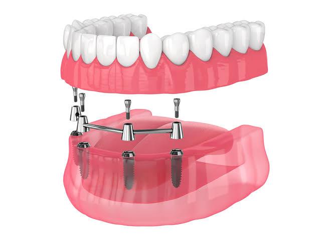
- Full-Arch Fixed Solutions: When an entire dental arch has failed due to advanced periodontal disease or extensive decay, full-arch restorations provide permanent stability. Using four to six strategically angled implants, an entire arch of teeth locks rigidly into place. Patients researching quality dental implants in san antonio frequently evaluate full-arch fixed bridges as an alternative to conventional dentures, specifically to avoid slipping appliances, messy adhesives, and dietary restrictions.
Surgical Phases and the Healing Horizon
The entire implant process is a multistep medical journey rather than a single office visit.
1. Site Preparation and Placement
The surgical site is numbed with local anesthesia, occasionally supplemented with oral or IV sedation for patient comfort. The dentist or oral surgeon creates a minor incision in the gingival tissue, prepares a precise osteotomy in the jawbone, and threads the titanium fixture into position. If primary stability is exceptionally high, a temporary crown may be placed; more frequently, a healing cap is attached, and the gum tissue is sutured closed over or around the site.
2. Healing and Osseointegration
During the next 12 to 24 weeks, the bone fuses with the implant. Patients generally experience mild soreness and minor swelling for only three to five days post-surgery, easily managed with over-the-counter anti-inflammatory medications and cold compresses. Hard, crunchy, or sticky foods must be avoided near the site to prevent micromovements that could disrupt bone remodeling.
3. Abutment Placement and Final Impression
Once digital imaging confirms bone consolidation, the fixture is exposed if covered by tissue, and an abutment is connected. A digital intraoral scanner captures the exact margins of the abutment, the gum contour, and the opposing bite.
4. Custom Crown Delivery
A dental laboratory mills a custom porcelain or zirconia restoration matched to the shade and translucency of the adjacent natural teeth. The crown is either screw-retained or cemented firmly into place, restoring full bite capability.
Protecting the Investment: Preventing Peri-Implantitis
While porcelain crowns will never develop cavities, the surrounding living bone and gingiva remain vulnerable to bacterial infections. The primary threat to a healed implant is peri-implantitis—an inflammatory condition triggered by bacterial biofilm that causes progressive bone loss around the titanium post.
Maintaining an implant requires daily mechanical hygiene:
- Clean around the abutment collar using non-abrasive fluoride toothpaste to prevent microscopic scratches where bacteria can lodge.
- Clean interdental spaces using unwaxed floss, specialized implant tape, or an interdental brush coated with plastic to avoid scratching the metal.
- Use a water flosser on low-to-medium pressure to flush food debris from deep margins around bridge connections.
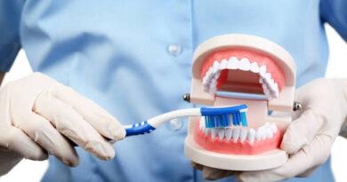
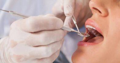
- Schedule professional cleanings every six months. Hygienists use resin, titanium, or carbon-fiber scalers rather than traditional stainless steel tools, preserving the smooth polish of the implant components.
Investing in permanent tooth replacement requires patience, thorough diagnostics, and consistent daily maintenance. When planned carefully around underlying bone biology, dental implants restore chewing force, preserve facial profile integrity, and deliver durable, natural function for decades.
Which stage of the tooth replacement process are you currently navigating—evaluating your initial candidacy or comparing specific restorative options for your smile?