
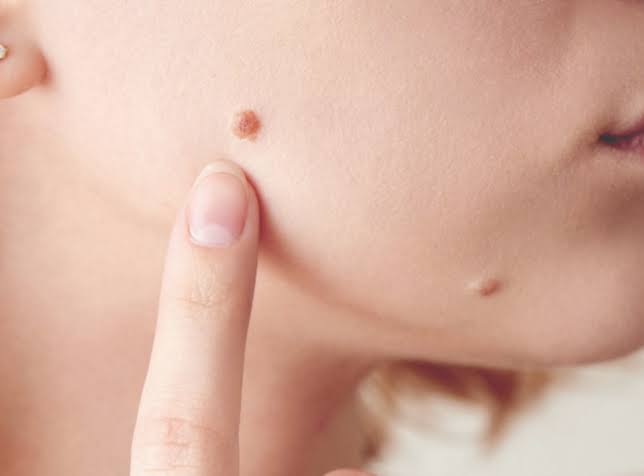
When May a Suspicious Mole Need a Skin Biopsy?
During a biopsy, part or all of the lesion is removed and sent to a pathology laboratory. Microscopic tissue examination provides the diagnosis, helping a doctor determine whether the mole is benign, atypical, melanoma, or another type of skin lesion.
Which Changes Can Make a Mole Suspicious?
A changing mole does not automatically mean cancer, but it should receive professional assessment. If a suspicious lesion requires further investigation, patients may naturally wonder whether a skin biopsy hurts before undergoing the procedure.
The ABCDE guide is useful, but melanomas do not always follow it, so any spot that looks different or behaves unusually deserves attention, particularly when the change is recent or continuing.
- Asymmetry: One half of the mole does not match the shape or structure of the other half.
- Border changes: The edge becomes irregular, blurred, scalloped, ragged, or difficult to define.
- Colour variation: Several shades appear within one lesion, including brown, black, red, blue, white, pink, or skin colour.
- Increasing diameter: The mole grows wider, although a small lesion can still be suspicious.
- Evolution: Its size, shape, colour, height, texture, or sensation changes over time.
- Bleeding or crusting: The area repeatedly bleeds, forms a crust, or develops a surface that does not heal.
- New sensation: Persistent itching, tenderness, irritation, or pain develops without an obvious injury.
- Different appearance: One mole looks noticeably unlike the person’s other spots, sometimes called the ugly duckling sign.
- Rapid growth: A raised, firm, or changing lesion develops over weeks or months.
- New mole later in life: A recently appearing spot may warrant closer assessment, especially when other suspicious features are present.
How Does a Doctor Decide Whether a Biopsy Is Needed?
A biopsy decision is based on more than one visible feature. The doctor considers the lesion, the patient’s history, dermoscopic findings, changes over time, and the consequences of monitoring an uncertain spot.
1. Reviews the Person’s Skin Cancer Risk
The doctor may ask about previous melanoma or other skin cancers, family history, severe sunburn, outdoor work, solarium use, immune suppression, and the number of unusual moles.
Risk does not determine the diagnosis. Personal history changes the level of concern, helping the doctor decide whether monitoring is reasonable or tissue examination is safer.
2. Examines the Mole With Dermoscopy
A dermatoscope magnifies the lesion and reveals structures beneath the outer skin surface. These patterns may help distinguish a typical mole from one needing further investigation.
Dermoscopy improves assessment when used by a trained clinician. It cannot replace pathology, so a lesion may still need biopsy when its appearance remains suspicious or uncertain.
3. Compares It With Other Moles
Many people have a recognisable pattern across their moles. A spot that differs in colour, structure, size, or behaviour may stand out even without meeting every ABCDE feature.
This comparison provides valuable context. A unique lesion may deserve closer attention, particularly when it is also new, growing, or changing.
4. Checks Whether the Lesion Is Evolving
Photographs, previous measurements, or the patient’s observations may show that a mole has changed. Evolution can be more informative than its appearance during one appointment.
A clinician may monitor selected lesions using sequential imaging. Documented change can trigger biopsy, even when the original image did not show definite signs of melanoma.
5. Considers the Lesion’s Location
Moles can occur on the scalp, soles, palms, beneath nails, and other areas that are difficult to monitor. Some locations also make complete removal or wound closure more complicated.
The doctor considers both diagnostic needs and procedure planning. Location can affect the biopsy method, referral decision, and specialist involvement.
6. Assesses Whether Monitoring Is Safe
Some atypical lesions can be photographed and reviewed after a planned interval. Monitoring may be unsuitable when melanoma is strongly suspected, follow-up is uncertain, or the lesion is changing quickly.
Patients should not choose their own monitoring schedule. Observation requires a clear clinical plan and prompt reassessment if changes appear before the booked review.
7. Uses Pathology to Resolve Uncertainty
A biopsy may be the only reliable way to determine what a suspicious mole contains. A pathologist examines the removed tissue and reports the cellular findings.
What Happens During a Skin Biopsy?
The procedure varies according to the lesion’s size, depth, location, and suspected diagnosis. The doctor should explain the proposed method, possible risks, wound care, and alternatives before requesting consent.
- The lesion and surrounding skin are examined and marked if necessary.
- The area is cleaned to reduce contamination risk.
- Local anaesthetic is injected to numb the procedure site.
- The doctor removes part or all of the suspicious lesion.
- An excision biopsy generally removes the complete lesion with a small surrounding margin.
- A partial biopsy may be selected for a large lesion or one in a difficult location.
- The wound may be closed with stitches, depending on its size and biopsy type.
- A dressing is placed over the area.
- The tissue sample is labelled and sent to a pathology laboratory.
- A pathologist examines the tissue under a microscope.
- The patient receives instructions about keeping the wound clean and dry.
- A follow-up appointment may be arranged for results and stitch removal.
- Further treatment is planned if the report identifies cancer or incomplete removal.
For a lesion suspected of being melanoma, complete excision biopsy is generally preferred when practical because it allows the pathologist to assess the entire lesion. The doctor may refer the patient to another GP, dermatologist, or surgeon when the site or procedure requires additional expertise.
What Should Patients Know After the Biopsy?
Recovery is usually straightforward, but good wound care and timely follow-up remain important. Patients should use their doctor’s instructions because biopsy sites and closure methods differ.
1. Follow the Wound-Care Instructions
Keep the dressing in place for the advised period and clean the area only as directed. Avoid applying unapproved creams, antiseptics, or home remedies.
Some soreness can occur after the anaesthetic wears off. Correct wound care supports healing and reduces avoidable irritation, bleeding, or infection.
2. Watch for Possible Complications
Contact the treating clinic if bleeding does not stop with advised pressure, pain becomes worse, the wound opens, or increasing redness and discharge develop.
A small scar is possible after any skin biopsy. Unexpected symptoms need professional advice, rather than repeated dressing changes without assessment.
3. Attend the Results Appointment
Do not assume that no news means a normal result. Confirm how and when the clinic will provide the pathology report before leaving the appointment.
The doctor needs to interpret the findings in context. Every biopsy result deserves clear communication, including whether the lesion was fully removed and whether further action is required.
4. Understand That Benign Results Are Common
Many biopsied moles prove to be benign or atypical rather than cancerous. The procedure may still have been appropriate because clinical examination could not provide certainty.
A benign result does not make the decision unnecessary. Biopsy is used to answer uncertainty, not because the doctor already knows that cancer is present.
5. Ask About Further Excision
If melanoma or another skin cancer is identified, further surgery may be recommended to remove additional surrounding tissue. The required treatment depends on the diagnosis and pathology features.
Further excision does not automatically mean the cancer has spread. Treatment decisions follow the pathology report, including information such as cancer type, depth, margins, and other relevant findings.
6. Continue Checking the Rest of the Skin
Removing one suspicious mole does not prevent another lesion from developing elsewhere. Continue regular self-checks and attend professional examinations according to medical advice.
Check areas that are easy to miss, including the scalp, back, soles, and nails. Ongoing awareness remains important after both benign and positive biopsy results.
7. Maintain Daily Sun Protection
A biopsy investigates an existing lesion but does not reverse earlier UV damage. Protective clothing, shade, broad-brimmed hats, sunglasses, and suitable sunscreen remain important.
Check the UV Index rather than relying on heat or cloud cover. Sun protection reduces future exposure, while skin checks from experts like skinchx.com.au support early recognition of new changes.
Conclusion
A suspicious mole may need biopsy when it changes, looks different from other spots, bleeds, grows, or shows concerning dermoscopic features. The procedure removes tissue for examination by a pathologist, providing a diagnosis that visual inspection alone cannot confirm. Follow wound-care advice, attend the results appointment, and seek prompt medical review for any new or changing skin lesion.